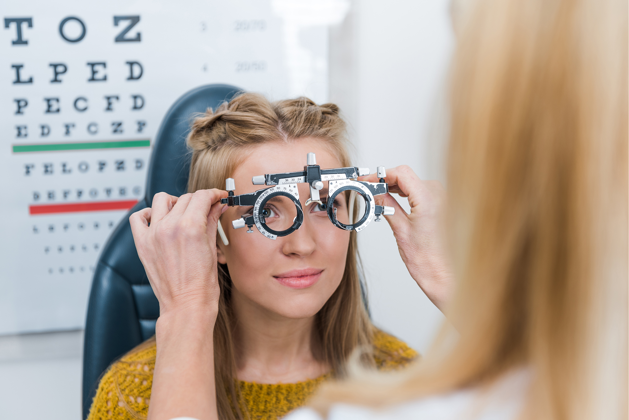
Eye Examinations
Comprehensive Eye Tests in Rayners Lane, Pinner – Eye Health Checks & Vision Screening
At Barnett Opticians, our thorough eye examinations go beyond simple vision checks. We screen for glaucoma, retinal health, and other conditions that affect long term eye health. Whether you need a routine eye test near you or have specific vision concerns, our experienced optometrists provide tailored care using the latest diagnostic technology.
Book your eye test online or call us today.
Book your eye test online or call us today.

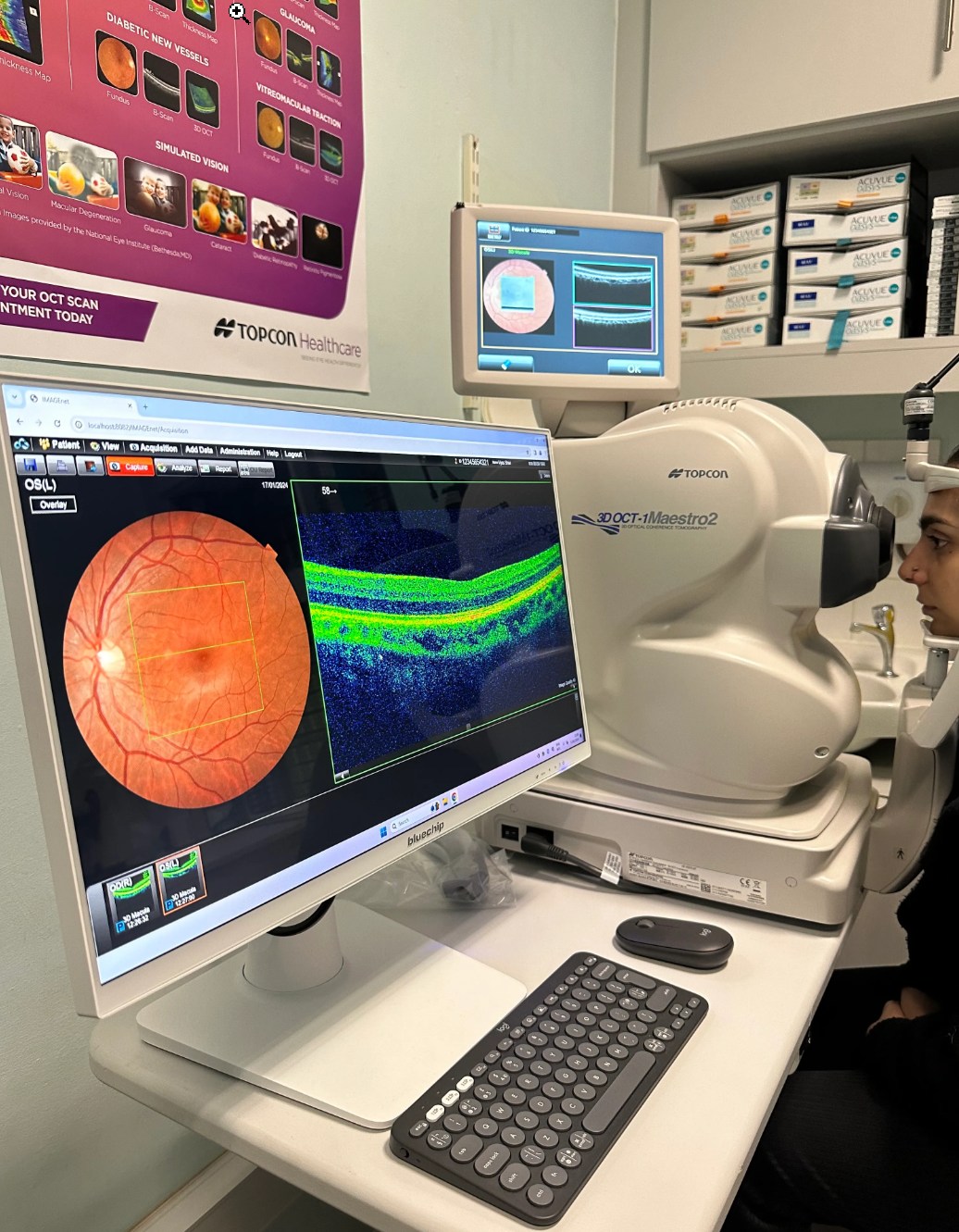
Enhanced Eyecare with OCT Technology
We use the latest technology to provide a thorough service. Optical Coherence Tomography (OCT) is a cutting-edge imaging technology that allows us to capture highly detailed, cross-sectional images of the retina. This advanced scan helps detect eye conditions such as glaucoma, macular degeneration, and diabetic retinopathy at their earliest stages, often before symptoms appear.
We highly recommend adding an OCT scan to your eye examination, whether you are a private or NHS patient, for a more thorough assessment of your eye health.
What Happens After Your Eye Examination?
After the eye examination your optometrist will tell you about the standard of your vision, the health of your eyes and any individual visual requirements you may have. You will be told whether spectacles are required and what to use them for. You will also be advised when your next eye examination is due.
If spectacles are required, you will see the registered dispensing optician who will advise on frame styles and lens types suitable for your lifestyle and visual needs.
If spectacles are required, you will see the registered dispensing optician who will advise on frame styles and lens types suitable for your lifestyle and visual needs.


Eyecare Plan – Comprehensive Care with Exclusive Benefits
Looking after your eyes has never been easier with our Eyecare Plan. Designed to provide complete peace of mind, our plan offers regular eye examinations, emergency appointments, and contact lens aftercare, ensuring your vision remains in top condition. As a member, you’ll also enjoy priority appointments, exclusive discounts on spectacles and accessories, and early access to the latest eyewear innovations.
Minor Eye Conditions Service (MECS)
We provide fast access to expert care for urgent but non-serious eye problems. If you experience symptoms such as red or painful eyes, sudden changes in vision, flashes or floaters, or ongoing eye discomfort, you can be assessed promptly by a qualified optometrist. MECS ensures you receive the right advice, treatment, or referral where needed, helping to resolve eye concerns quickly and safely without unnecessary delays.